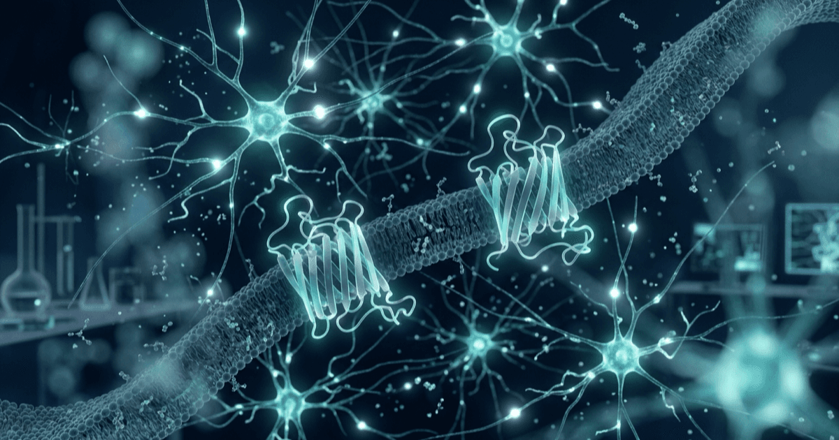
Published by CertaPeptides. For research purposes only.
The hypothalamus contains a small number of neuron populations that set the tempo of reproductive neuroendocrine signalling. Two peptide systems come up again and again in that literature: kisspeptin, acting through the receptor KISS1R (also called GPR54), and oxytocin, acting through the oxytocin receptor (OXTR). Both are G protein-coupled receptor (GPCR) systems, both are expressed in hypothalamic circuits, and both have become standard tools for probing how peptide signals control neuronal excitability and hormone release in cell and animal models.
This guide gives an overview of the research category behind our gonadotropic research peptides. It covers the pathway as a field of study, how kisspeptin-10 and oxytocin are used as receptor probes, where the two systems meet, and the general laboratory practices that apply to lyophilised peptides of this kind. For a deeper discussion of kisspeptin specifically, see our dedicated article on kisspeptin-10 in reproductive neuroendocrine research.
What is the gonadotropic pathway as a research field?
In vertebrate physiology, the term “gonadotropic pathway” describes the chain of signals that links the brain to the gonads, often called the hypothalamic-pituitary-gonadal (HPG) axis. At its top sits a scattered population of hypothalamic neurons that secrete gonadotropin-releasing hormone (GnRH) into the portal blood of the median eminence. GnRH acts on pituitary gonadotrope cells, which in turn release luteinising hormone (LH) and follicle-stimulating hormone (FSH). In laboratory work these endogenous hormones are mostly measured as read-outs: a rise or fall in LH in an animal model is used as a proxy for what GnRH neurons are doing.
The research questions in this field are largely neurobiological. How do GnRH neurons generate pulses? Which upstream neurons drive them? How do steroid hormones feed back onto the hypothalamus? Which receptors and second-messenger pathways translate a peptide signal into a change in firing? Answering these questions needs selective ligands that act on defined receptors, which is where research peptides such as kisspeptin-10 and oxytocin come in.
Why GPCR signalling matters here
Most of the neuropeptide receptors in this system belong to the class A (rhodopsin-like) GPCR family. When an agonist binds, the receptor changes shape and engages heterotrimeric G proteins. For both KISS1R and OXTR, the main coupling studied is to Gq/11 proteins, which activate phospholipase C-beta (PLC-beta). PLC-beta hydrolyses the membrane lipid PIP2 into inositol trisphosphate (IP3) and diacylglycerol. IP3 releases calcium from intracellular stores, and diacylglycerol activates protein kinase C. Downstream, research groups track calcium transients, MAP kinase phosphorylation (ERK1/2, p38) and changes in membrane potential as measurable outputs.
Because these read-outs can be measured in transfected cell lines, in brain slices and in whole animals, the same ligand can be followed from receptor binding through to circuit-level effects. That continuity is a large part of why kisspeptin and oxytocin are so widely used as research tools.
Kisspeptin-10: a KISS1R/GPR54 agonist research tool
This section is a short summary. Our kisspeptin-10 reference article covers receptor signalling and HPG axis research in more depth.
Discovery and receptor pharmacology
Kisspeptins were identified in 2001 by two independent groups as the endogenous ligands of the then-orphan receptor GPR54. Ohtaki and colleagues isolated a 54-residue C-terminally amidated peptide from human placenta, encoded by the KiSS-1 gene, and named it metastin because of the gene’s earlier association with metastasis suppression in cell models [2]. Kotani and colleagues independently isolated 54-, 14- and 13-residue peptides sharing a common RF-amide C terminus and named them kisspeptins [1].
Kisspeptin-10 is the C-terminal decapeptide shared by these longer forms (sequence Tyr-Asn-Trp-Asn-Ser-Phe-Gly-Leu-Arg-Phe-NH2). In the receptor characterisation work, kisspeptins bound rat and human GPR54 expressed in Chinese hamster ovary cells with low nanomolar affinity, and receptor activation stimulated PIP2 hydrolysis, calcium mobilisation, arachidonic acid release, ERK1/2 and p38 MAP kinase phosphorylation and stress fibre formation [1]. This profile is typical of a Gq/11-coupled receptor, and it is the basis for using kisspeptin-10 as a defined agonist in in vitro assays of KISS1R signalling.
Kisspeptin as a probe of GnRH-neuron activation
The most widely cited animal-model work established that kisspeptin acts at the level of the GnRH neuron itself. In mice, GPR54 transcripts were found to colocalise with GnRH neurons; kisspeptin raised LH and FSH in wild-type animals but had no effect in mice lacking Gpr54; and in sheep, central kisspeptin produced a marked release of GnRH into cerebrospinal fluid, measured directly [3]. Together these observations positioned GPR54 as a key control point in the reproductive axis and kisspeptin as a direct GnRH-releasing signal in these models.
Electrophysiology in brain slices then showed what happens at the cellular level. Using GnRH-GFP mice, Han and colleagues reported that kisspeptin evoked a potent, long-lasting depolarisation of more than 90% of adult GnRH neurons, while a much smaller fraction of neurons responded in juvenile and prepubertal animals [4]. Around 90% of GnRH neurons expressed GPR54 mRNA at both ages, which led the authors to propose that the developmental change reflects both increased kisspeptin input and a post-transcriptional change in receptor signalling within the GnRH neuron [4].
In practice, this means researchers use kisspeptin-10 in three main ways:
- as a reference agonist in cell-based KISS1R assays (calcium imaging, IP accumulation, kinase phosphorylation);
- as a stimulus in slice electrophysiology to test whether identified GnRH neurons are responsive, and how that responsiveness changes with developmental stage or steroid environment;
- as a probe in animal models, where an LH response is used as a functional read-out of an intact kisspeptin-GnRH connection.
KNDy neurons: the upstream pulse generator
Much current work focuses on where endogenous kisspeptin comes from. In the arcuate nucleus, a population of neurons co-expresses kisspeptin, neurokinin B and dynorphin, giving them the name KNDy neurons. Navarro and colleagues showed by in situ hybridisation that arcuate Kiss1 neurons in female mice also express the genes for dynorphin and neurokinin B, as well as their receptors (the kappa opioid receptor and NK3), and that this expression is inhibited by estradiol [5]. They proposed a model in which neurokinin B and dynorphin act on the kisspeptin neurons themselves to synchronise and shape pulsatile kisspeptin output, which then drives GnRH release from fibres in the median eminence [5].
A widely cited review by Lehman, Coolen and Goodman describes KNDy cells as a central node in the control of GnRH secretion, drawing together anatomical and functional data from several species [6]. For researchers, the KNDy model explains why exogenous kisspeptin-10 is so useful: it lets an experiment bypass the upstream pulse generator and test the downstream GnRH neuron directly.
Oxytocin: an OXTR research ligand
Oxytocin is a nine-residue peptide (Cys-Tyr-Ile-Gln-Asn-Cys-Pro-Leu-Gly-NH2) with a disulfide bridge between the two cysteine residues that closes a six-residue ring, and an amidated C terminus. It belongs to the neurohypophysial peptide family together with vasopressin, from which it differs by two residues. That close structural relationship is important experimentally, because oxytocin and vasopressin receptors form a related family and cross-reactivity has to be considered when interpreting results [7].
Receptor structure and signalling
The oxytocin receptor is a typical class A GPCR that couples mainly through Gq proteins to PLC-beta [7]. Two allosteric factors stand out in the literature. First, the high-affinity receptor state depends on both magnesium ions and membrane cholesterol [7]. Second, these dependencies now have a structural explanation: the crystal structure of the human oxytocin receptor, solved in complex with a non-peptide antagonist, revealed a cholesterol molecule bound in an unexpected position between transmembrane helices IV and V and identified a conserved coordination site for Mg2+ that acts as a positive allosteric modulator of agonist binding [8].
For assay design, these findings have direct practical consequences. Membrane preparations or cell lines with altered cholesterol content, and buffers without divalent cations, can shift apparent oxytocin affinity. Studies indicate that controlling these variables is part of producing reproducible OXTR binding data [7, 8].
Beyond Gq, the receptor’s signalling repertoire is broader than a single pathway. A review by Busnelli and Chini summarises how OXTR couples to different G proteins, which signalling cascades it activates in neuronal and glial cells, and how receptor trafficking at the plasma membrane and inside the cell shapes the size and persistence of the response [9].
Receptor desensitisation and trafficking
Like many GPCRs, OXTR does not respond indefinitely to continued agonist exposure. After activation, receptors are phosphorylated, recruit arrestin proteins and are internalised, after which they can be recycled to the membrane or degraded. Reviews of the system describe how this regulation, together with steroid-dependent changes in receptor expression, controls how strongly a given tissue responds to oxytocin [7, 9]. In cell-based work this matters for experimental design: repeated or prolonged agonist exposure can reduce the measured response, so time-course controls and receptor-expression measurements are often reported alongside functional read-outs.
Hypothalamic magnocellular neurons
The main site of oxytocin gene expression is the population of magnocellular neurons in the paraventricular and supraoptic nuclei of the hypothalamus. These neurons project to the posterior pituitary, where oxytocin is released into the circulation, but they also release oxytocin within the hypothalamic nuclei themselves, and oxytocin neurons project widely through the central nervous system [7].
A notable feature of these cells is dendritic release. Ludwig and Leng described how neuropeptides such as oxytocin and vasopressin are released from dendrites, acting as autocrine or paracrine signals at their site of origin, and how activity-dependent priming of dendritic vesicle stores could reorganise local networks for extended periods [10]. This makes the magnocellular system a model of choice for studying how a single peptide can act both as a circulating hormone and as a local neuromodulator.
In vitro and animal-model research
Oxytocin is used as a reference agonist in a range of experimental settings:
- receptor binding and functional assays in cell lines expressing recombinant OXTR, including studies of allosteric modulation by Mg2+ and cholesterol;
- calcium imaging and second-messenger assays to characterise Gq-dependent signalling;
- trafficking studies following receptor internalisation and recycling;
- slice and in vivo electrophysiology of supraoptic and paraventricular neurons;
- tissue and organ-bath preparations in which receptor-expressing smooth muscle is used as a functional assay in animal tissue.
Reviews also describe oxytocin receptor expression in peripheral tissues such as kidney, heart, thymus, pancreas and adipocytes in animal studies, which gives researchers a range of tissues in which to compare receptor regulation [7].
Researchers can find the peptide on our oxytocin acetate product page.
Where the kisspeptin and oxytocin systems intersect
The two systems are often studied separately, but there are several points of overlap that make them useful to consider together.
Kisspeptin and supraoptic oxytocin neurons
The most direct link comes from electrophysiology. Scott and Brown recorded single supraoptic neurons in anaesthetised female rats and found that kisspeptin-10 acting in the periphery increased the firing rate of oxytocin neurons, whereas only a minority of vasopressin neurons responded, with short high-frequency bursts [11]. Kisspeptin-10 delivered centrally did not alter firing. When vagal afferents were desensitised, the peripheral effect on oxytocin neurons was prevented, suggesting that kisspeptin reached the supraoptic nucleus through vagal afferent input, acting as a hormone rather than as a local neuropeptide in this system [11].
This finding is a good example of why the site of action and circuit context matter when interpreting research-ligand data: the same peptide produced different results depending on whether it acted inside or outside the brain in this rat model.
Shared signalling logic
Both KISS1R and OXTR are Gq-coupled GPCRs on hypothalamic neurons, and both produce calcium-dependent changes in excitability [1, 7]. This shared biochemistry means many of the same assay platforms (calcium imaging, IP accumulation, kinase phosphorylation, patch-clamp recording) can be used for both, which simplifies comparative studies.
Steroid sensitivity
Both systems are strongly shaped by gonadal steroids. Arcuate Kiss1, dynorphin and neurokinin B expression is inhibited by estradiol in mice [5], and the regulation of the oxytocin system by gonadal and adrenal steroids is described as one of its most remarkable, and least understood, features [7]. Experiments that compare responsiveness across steroid conditions are therefore common in both fields, and controlling the endocrine state of animal models is a standard design consideration.
Peptide release from hypothalamic neurons
Finally, both systems rely on neurosecretory release from hypothalamic neurons: kisspeptin from KNDy and other kisspeptin populations onto GnRH neurons and their terminals [5, 6], and oxytocin from magnocellular neurons both to the pituitary and locally through dendrites [7, 10]. Studies that ask how peptide release is coordinated across neuron populations draw on both literatures.
Laboratory considerations for lyophilised peptides
The points below describe general laboratory practice for short synthetic peptides of this type. They are not specific instructions, and each laboratory should follow its own standard operating procedures and safety rules.
Storage
Lyophilised (freeze-dried) peptides are generally stored at -20°C, protected from light and moisture, in a sealed container with desiccant where possible. Before opening, letting the vial reach room temperature reduces condensation on the dry powder. Our peptide storage guide covers temperature, light and humidity in more detail, and our article on peptide lyophilisation explains why the freeze-dried state is the most stable form for storage.
Reconstitution as laboratory chemistry
Reconstitution means dissolving the lyophilised solid in a suitable solvent to make a stock solution of known concentration for in vitro or analytical work. The variables are the solvent, the volume and the resulting concentration:
- Solvent. Many short peptides dissolve in sterile, high-purity water. Oxytocin is generally water-soluble. Kisspeptin-10 contains several aromatic and hydrophobic residues, so some laboratories use a small proportion of a compatible co-solvent, or a dilute aqueous acid, before diluting into an assay buffer. The solvent should be compatible with the downstream assay.
- Volume and concentration. Stock concentration is the mass of peptide divided by the volume of solvent added. Laboratories usually record the calculation on the tube label and in the lab notebook, and correct for net peptide content if that information is available.
- Technique. Gentle swirling rather than vigorous shaking limits foaming and surface adsorption. Low-binding tubes can reduce peptide loss for dilute solutions.
Handling and stability in solution
Peptides in solution are less stable than the dry powder. Common practice is to divide a stock into single-use aliquots, store them frozen, and avoid repeated freezing and thawing. Oxytocin’s disulfide bond and kisspeptin-10’s tryptophan residue can both be sensitive to oxidation, so limiting air exposure and light is reasonable. Working dilutions are typically prepared fresh on the day of the experiment.
Analytical identity confirmation
As a general laboratory practice, many research groups confirm the identity and integrity of a peptide before starting a study, for example by chromatographic purity assessment and mass-based identity confirmation in their own facility or a contract laboratory. This is part of good experimental hygiene and helps rule out degradation as a cause of unexpected results. Product pages show a class-level specification where one is on record. For kisspeptin-10, reports for selected lots independently tested are searchable by batch or report code at certapeptides.com/verify.
Kisspeptin-10 is available on our kisspeptin-10 product page, and both peptides are listed in the gonadotropic research category.
Key takeaways
- The gonadotropic pathway, or HPG axis, is studied mainly as a neurobiological question: how hypothalamic neurons generate and regulate GnRH output, measured through pituitary hormone read-outs in models.
- Kisspeptin-10 is the C-terminal decapeptide of the kisspeptins, the endogenous ligands of KISS1R/GPR54, a Gq/11-coupled receptor [1, 2].
- Animal-model and slice studies indicate that kisspeptin acts directly on GnRH neurons through GPR54 [3, 4], which makes kisspeptin-10 a standard probe of GnRH-neuron activation.
- Arcuate KNDy neurons are thought to act as the upstream pulse generator that supplies endogenous kisspeptin [5, 6].
- Oxytocin is a cyclic nonapeptide that acts on OXTR, a Gq-coupled GPCR whose high-affinity state depends on Mg2+ and cholesterol [7, 8]; receptor trafficking and desensitisation shape response size and duration [9].
- Magnocellular oxytocin neurons release peptide both to the pituitary and locally from dendrites [7, 10].
- The systems intersect: in a rat model, kisspeptin-10 acting peripherally increased supraoptic oxytocin-neuron firing through vagal afferents [11].
- Lyophilised peptides are generally stored at -20°C; reconstitution is a matter of solvent, volume and concentration; aliquoting and identity confirmation are good general practice.
Frequently asked questions
What does “gonadotropic research” cover?
It covers laboratory research on the hypothalamic-pituitary-gonadal axis, particularly the hypothalamic neurons and receptors that control GnRH and pituitary hormone release. Typical work includes receptor pharmacology in cell lines, slice electrophysiology and animal-model studies in which hormone levels are used as read-outs.
Which receptor does kisspeptin-10 act on?
Kisspeptin-10 is an agonist of KISS1R, originally described as the orphan receptor GPR54. In cell-based studies, receptor activation stimulated PIP2 hydrolysis, calcium mobilisation and MAP kinase phosphorylation, consistent with Gq/11 coupling [1].
How does kisspeptin-10 differ from longer kisspeptins?
The longer endogenous forms (54, 14 and 13 residues) share the same RF-amide C-terminal sequence [1, 2]. Kisspeptin-10 is that shared C-terminal decapeptide, which makes it a convenient, short synthetic agonist for receptor studies.
What are KNDy neurons?
KNDy neurons are arcuate nucleus neurons that co-express kisspeptin, neurokinin B and dynorphin. Research suggests they generate the pulsatile kisspeptin signal that drives GnRH release in rodent and other animal models [5, 6]. Our kisspeptin-10 article discusses them further.
How does the oxytocin receptor signal?
OXTR is a class A GPCR that couples mainly through Gq proteins to phospholipase C-beta, raising intracellular calcium [7]. Its high-affinity state depends on Mg2+ and cholesterol, both of which are explained by the receptor’s crystal structure [8]. It can also engage other signalling pathways and is regulated by internalisation and recycling [9].
Is there evidence that kisspeptin affects oxytocin neurons?
In anaesthetised female rats, kisspeptin-10 acting peripherally increased the firing of supraoptic oxytocin neurons, an effect prevented by desensitising vagal afferents; central kisspeptin-10 did not alter firing in that study [11].
How should lyophilised peptides be stored in the lab?
As a general rule, at -20°C, dry, protected from light and in a sealed container. Once reconstituted, solutions are usually aliquoted and frozen to avoid repeated freezing and thawing. See our storage guide for details.
References
- Kotani M, Detheux M, Vandenbogaerde A, et al. The metastasis suppressor gene KiSS-1 encodes kisspeptins, the natural ligands of the orphan G protein-coupled receptor GPR54. J Biol Chem. 2001;276(37):34631-34636. doi:10.1074/jbc.M104847200
- Ohtaki T, Shintani Y, Honda S, et al. Metastasis suppressor gene KiSS-1 encodes peptide ligand of a G-protein-coupled receptor. Nature. 2001;411(6837):613-617. doi:10.1038/35079135
- Messager S, Chatzidaki EE, Ma D, et al. Kisspeptin directly stimulates gonadotropin-releasing hormone release via G protein-coupled receptor 54. Proc Natl Acad Sci U S A. 2005;102(5):1761-1766. doi:10.1073/pnas.0409330102
- Han SK, Gottsch ML, Lee KJ, et al. Activation of gonadotropin-releasing hormone neurons by kisspeptin as a neuroendocrine switch for the onset of puberty. J Neurosci. 2005;25(49):11349-11356. doi:10.1523/JNEUROSCI.3328-05.2005
- Navarro VM, Gottsch ML, Chavkin C, et al. Regulation of gonadotropin-releasing hormone secretion by kisspeptin/dynorphin/neurokinin B neurons in the arcuate nucleus of the mouse. J Neurosci. 2009;29(38):11859-11866. doi:10.1523/JNEUROSCI.1569-09.2009
- Lehman MN, Coolen LM, Goodman RL. Minireview: kisspeptin/neurokinin B/dynorphin (KNDy) cells of the arcuate nucleus: a central node in the control of gonadotropin-releasing hormone secretion. Endocrinology. 2010;151(8):3479-3489. doi:10.1210/en.2010-0022
- Gimpl G, Fahrenholz F. The oxytocin receptor system: structure, function, and regulation. Physiol Rev. 2001;81(2):629-683. doi:10.1152/physrev.2001.81.2.629
- Waltenspühl Y, Schöppe J, Ehrenmann J, Kummer L, Plückthun A. Crystal structure of the human oxytocin receptor. Sci Adv. 2020;6(29):eabb5419. doi:10.1126/sciadv.abb5419
- Busnelli M, Chini B. Molecular basis of oxytocin receptor signalling in the brain: what we know and what we need to know. Curr Top Behav Neurosci. 2018;35:3-29. doi:10.1007/7854_2017_6
- Ludwig M, Leng G. Dendritic peptide release and peptide-dependent behaviours. Nat Rev Neurosci. 2006;7(2):126-136. doi:10.1038/nrn1845
- Scott V, Brown CH. Kisspeptin activation of supraoptic nucleus neurons in vivo. Endocrinology. 2011;152(10):3862-3870. doi:10.1210/en.2011-1181
Disclaimer
For research purposes only. This article is educational; the compounds discussed are sold for laboratory research use only and are not for human consumption.
Ověřte si to před výzkumem
Stránky produktů uvádějí specifikaci na úrovni třídy, pokud je nějaká evidována, a vybrané šarže nesou nezávislý COA od třetí strany.