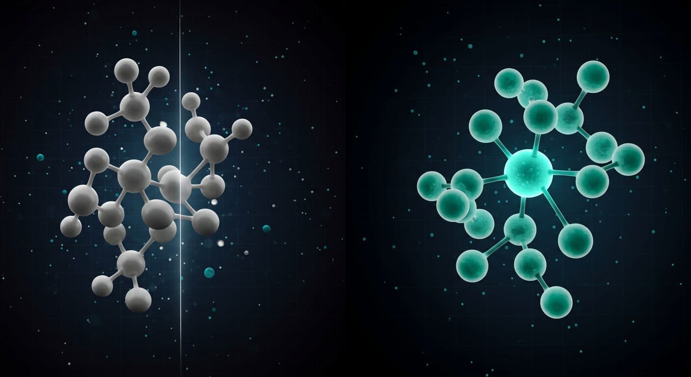
The distinction between GHK and GHK-Cu is not a marketing technicality — it is a structural and functional difference that materially affects experimental outcomes. Yet the two are conflated in product descriptions, research forums, and even occasional review articles. This post lays out what the published evidence actually says about how GHK and GHK-Cu differ: in stability, biological activity, and research application. If you are designing a study, sourcing a compound, or interpreting the literature, this distinction matters. This content is for educational and research purposes only.
The Chemistry First
GHK is a tripeptide: glycine-histidine-lysine (Gly-His-Lys). Molecular weight approximately 340 Da as the free peptide. It is found endogenously in human plasma, saliva, and urine, where it exists in both free form and as the copper chelate depending on copper availability.
GHK-Cu is GHK with a single Cu(II) ion chelated at two coordination sites: the imidazole nitrogen of the histidine residue and the alpha-amino nitrogen of the N-terminal glycine. The copper ion is not loosely associated — it forms a square-planar complex with a dissociation constant in the nanomolar range. This binding geometry is characteristic of type 2 copper sites in biology, similar to the copper-binding domain of ceruloplasmin.
Key structural consequences of copper chelation:
- Charge and polarity change: Cu(II) coordination alters the electronic structure of the histidine ring and the terminal amine, changing how the molecule interacts with cell surface receptors and plasma proteins
- Color: GHK is colorless in solution; GHK-Cu has a characteristic blue-green color at research concentrations due to Cu(II) d-d electronic transitions
- Redox activity: The copper ion is redox-active (Cu(II)/Cu(I) cycling), contributing to antioxidant and catalytic activities absent in GHK alone
- Molecular weight: GHK free peptide ~340 Da; GHK-Cu complex ~340.6 Da as the free complex (the copper contribution is approximately 63.5 Da, offset by proton displacement from the ligand)
For a complete overview of GHK-Cu’s molecular structure and research applications, see: GHK-Cu Copper Peptide: The Complete Research Guide.
Stability Differences
GHK (without copper) is a simple tripeptide with modest plasma stability. Tripeptides are cleaved by dipeptidyl peptidases and aminopeptidases that are abundant in plasma. Published data on GHK half-life in plasma is limited, but based on the structural properties of small peptides (no D-amino acids, no N-methylation, no other stability-enhancing modifications), rapid proteolysis is expected in vivo.
GHK-Cu shows meaningfully improved stability in plasma. The copper chelation geometry creates steric hindrance around the scissile peptide bonds, reducing protease accessibility. Additionally, the copper-bound form may interact with copper transport proteins (ceruloplasmin, albumin) that effectively chaperone the complex away from proteolytic enzymes.
Pickart’s early work noted that GHK-Cu appeared in the plasma albumin fraction — the same fraction that led to its discovery — suggesting that the copper chelate form has a preferred association with albumin that the free peptide lacks. This protein association is relevant to both plasma half-life and cellular uptake kinetics.
For research applications, this means:
- In vivo rodent studies where systemic exposure is relevant should use GHK-Cu, not GHK alone
- In long-duration cell culture experiments (24-72+ hours), GHK-Cu will likely maintain higher effective concentrations than GHK due to reduced proteolytic degradation
- For short-duration in vitro experiments (2-4 hours), the stability difference may be less important
Biological Activity Comparison in Published Studies
The most important direct comparison between GHK and GHK-Cu comes from the Maquart group’s fibroblast studies. Maquart et al. (1988) (PMID: 3169264) ran parallel experiments with GHK and the copper complex in cultured human skin fibroblasts, measuring collagen synthesis by radiolabeled proline incorporation and consistently finding greater activity with the copper-chelated form.
Their findings consistently showed that GHK-Cu produced greater stimulation of both collagen and glycosaminoglycan synthesis than GHK alone at equivalent molar concentrations. The effect was not marginal — GHK showed modest activity while GHK-Cu showed substantially greater activity at the same concentrations. This study is the primary direct evidence that copper chelation is required for the full biological activity of this compound class.
Possible explanations the authors proposed:
- The copper ion participates directly in receptor binding, not just as a structural modifier
- Copper provides a local source for lysyl oxidase (LOX) activation, augmenting the collagen synthesis effect with enhanced crosslinking capability
- The changed charge state of GHK-Cu vs. GHK affects cellular uptake kinetics
None of these explanations has been definitively established at the molecular level, but the empirical observation of differential activity is well-documented.
What the Original Research Actually Studied
This is a critical point for literature interpretation. When you read a paper citing “GHK-Cu” effects, you need to verify what compound was actually used. The literature contains three categories:
Studies using GHK-Cu (the copper chelate)
Most of the wound healing and hair follicle literature from Pickart’s lab used the copper chelate. The characteristic blue-green solution color in published photographs confirms copper chelation.
Studies using GHK (the free tripeptide)
Some more recent studies, particularly gene expression analyses, have used GHK (without copper) as a stimulus. Pickart, Vasquez-Soltero, and Margolina’s (2015) large-scale gene expression review (DOI: 10.1155/2015/648108) analyzed microarray datasets in which GHK (without copper) was the stimulus, and the authors explicitly discuss whether these findings extrapolate to the GHK-Cu complex. The honest answer is: partially, but not entirely — the copper ion is load-bearing for several activities in the fibroblast literature, so gene expression data on GHK alone should not be read as a direct proxy for GHK-Cu.
Cosmetic formulation studies
Clinical cosmetic studies (e.g., Fitzpatrick and Rostan, 2003 — DOI: 10.1080/14764170310000817) used complex formulations containing copper peptides but with variable specification of the exact chemical form. For interpreting cosmetic formulation outcomes, distinguish between “copper peptide formulation” studies and “purified GHK-Cu” studies.
Receptor and Uptake Mechanisms
How do GHK and GHK-Cu enter cells and interact with receptors? This is an area of active, incomplete research.
For copper uptake, the copper transporter 1 (CTR1, also called SLC31A1) is the primary mammalian copper importer. CTR1 preferentially transports Cu(I), which means Cu(II) in GHK-Cu must be reduced before or during transport. Whether the peptide scaffold facilitates this reduction or is cleaved prior to copper transport is not fully characterized.
Kang et al. (2009) (DOI: 10.1007/s00403-009-0942-x) specifically showed Copper–GHK upregulation of integrin α6 and β1 — the collagen- and laminin-binding integrin subunits — in human keratinocytes, along with increased PCNA and p63 positivity in monolayer and skin-equivalent culture. Whether this effect is replicated with GHK (without copper) was not examined in that study, leaving the question of copper requirement for integrin modulation open.
Practical Implications for Research Design
Which compound to use
- Use GHK-Cu for wound healing, collagen synthesis, angiogenesis, or hair follicle endpoints — these are the areas where the copper chelate shows greater activity in published comparisons
- GHK alone may be appropriate if you are specifically studying the peptide’s interaction with gene expression systems where copper’s redox activity would confound the interpretation
- Matched comparison groups (GHK vs. GHK-Cu vs. vehicle) are the cleanest design for distinguishing peptide from copper effects
Controls
Consider including a CuCl₂ control at equivalent copper concentrations to distinguish GHK-Cu-specific effects from non-specific copper effects. Several published studies have done this; those that have not leave the copper contribution ambiguous.
Concentration ranges
Published fibroblast studies have used GHK-Cu in the 0.1 nM to 10 nM range for gene expression effects and up to micromolar ranges for some cell culture endpoints. Because the copper chelate is more stable, effective concentrations at the receptor may be more predictable over time than with the free peptide.
Sourcing the Right Compound
When sourcing research material, confirm that the compound is GHK-Cu (the copper chelate), not GHK (the free tripeptide). A blue-green color in solution is a practical indicator of copper chelation. The COA should specify the molecular formula including copper (C₁₄H₂₂CuN₆O₄) and ideally include mass spectrometry data confirming the copper adduct ion.
Research-grade GHK-Cu is available at certapeptides.com/shop/ghk-cu. Our COA page (certapeptides.com/coa) shows a card for each tested GHK-Cu size; the Janoshik report on the 100mg lot (#212150) also states the measured copper content.
Key Takeaways
- GHK (free tripeptide) and GHK-Cu (copper chelate) are structurally distinct compounds with different plasma stability, cellular uptake kinetics, and biological activity profiles
- Direct comparisons in fibroblast studies (Maquart et al., 1993) consistently show greater collagen and glycosaminoglycan synthesis stimulation with GHK-Cu than GHK alone at equivalent molar concentrations
- The copper component contributes independently through lysyl oxidase cofactor activity — the peptide stimulates collagen synthesis while the copper facilitates collagen crosslinking
- Much of the published literature uses GHK-Cu; gene expression studies sometimes use GHK alone; always verify which compound was actually used before citing a study
- Treat a blue-green solution color only as a preliminary sign of copper chelation; where a report gives an analytical result for the copper-bound complex, check that result
References
- Kang YA, Choi HR, Na JI, et al. (2009). Copper–GHK increases integrin expression and p63 positivity by keratinocytes. Archives of Dermatological Research, 301(4), 301–306. DOI: 10.1007/s00403-009-0942-x
- Fitzpatrick RE, Rostan EF. (2003). Reversal of photodamage with topical growth factors. Journal of Cosmetic and Laser Therapy, 5(1), 25–34. DOI: 10.1080/14764170310000817
- Pickart L, Vasquez-Soltero JM, Margolina A. (2015). GHK Peptide as a Natural Modulator of Multiple Cellular Pathways in Skin Regeneration. BioMed Research International, 2015, Article 648108. DOI: 10.1155/2015/648108
- Pickart L, Margolina A. (2018). Regenerative and protective actions of the GHK-Cu peptide in the light of the new gene data. Biomolecules, 8(2), 29. DOI: 10.3390/biom8020029
- Gorouhi F, Maibach HI. (2009). Role of topical peptides in preventing or treating aged skin. International Journal of Cosmetic Science, 31(5), 327-345. DOI: 10.1111/j.1468-2494.2009.00490.x
References
- Pickart L, Thaler MM. (1973). Tripeptide in human serum which prolongs survival of normal liver cells and stimulates growth of neoplastic liver cells. Nature New Biology, 243(124), 85–87. PMID: 4349963.
- Maquart FX, et al. (1988). Stimulation of collagen synthesis in fibroblast cultures by the tripeptide-copper complex glycyl-L-histidyl-L-lysine-Cu2+. FEBS Letters, 238(2), 343–346. PMID: 3169264.
- Pickart L, Vasquez-Soltero JM, Margolina A. (2015). GHK Peptide as a Natural Modulator of Multiple Cellular Pathways in Skin Regeneration. BioMed Research International, 2015, 648108. PMID: 26236730.
All products are intended for research purposes only. Not for human consumption.
Verify before you research
Product pages show a class-level specification where one is on record, and selected lots carry an independent third-party COA.