⚠️ For Research Purposes Only — This article discusses MGF (mechano growth factor) as a laboratory research compound. The content is intended for scientific and educational review only. The peptide is not for human consumption and not intended to diagnose, treat, cure, or prevent any disease. All studies referenced describe preclinical investigations in cell culture or animal models.
Introduction
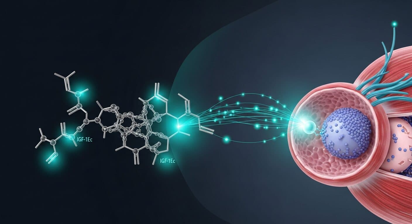
Mechano Growth Factor (MGF) is the name commonly given to a synthetic peptide corresponding to the 24-amino-acid C-terminal “E-domain” encoded by a mechanical load-responsive alternative splice variant of the insulin-like growth factor 1 (IGF1) gene. The mRNA splice variant itself is known as IGF-1Ec in humans (and IGF-1Eb in rodents, which has a 52-bp insert versus the 49-bp human insert). Researchers have named both the mRNA variant and the derived E-domain peptide “MGF” in the muscle physiology literature, though this dual usage is a common source of confusion.
According to PubMed, the field’s interest in MGF stems from observations that skeletal muscle IGF-1 mRNA undergoes alternative splicing in response to mechanical overload and injury, and that the IGF-1Ec/Eb transcript is upregulated early in this response, before later upregulation of the conventional IGF-1Ea isoform (Hill and Goldspink 2003, DOI; Matheny et al. 2010, DOI).
This article reviews the molecular biology of the IGF-1 splice variants that give rise to MGF, the hypothesized mechanisms by which the synthetic MGF E-peptide acts in research models, key findings in muscle regeneration and aging research, and crucially the published controversies over the reproducibility of MGF peptide effects in myoblasts and primary muscle stem cells.
Molecular Structure and Biochemistry
IGF-1 Gene Architecture
The IGF-1 gene is complex. Alternative splicing of exons 4, 5, and 6 generates multiple pro-IGF-1 mRNA variants that share the mature IGF-1 peptide but differ in their E-peptide (extension) regions. In humans, the main isoforms are IGF-1Ea (expressed constitutively and in systemic IGF-1), IGF-1Eb, and IGF-1Ec (upregulated by mechanical stress and injury in muscle). In rodents, the homolog of human IGF-1Ec is called IGF-1Eb. This nomenclature difference is important when reading the primary literature (Matheny et al. 2010, DOI; Dai et al. 2010, DOI).
The MGF E-Peptide
The synthetic peptide commonly sold and studied as “MGF” corresponds to the 24-amino-acid C-terminus of the unprocessed IGF-1Ec/Eb pro-IGF-1 molecule. This E-peptide region arises from the frame shift introduced by the alternative splice insert and terminates at a unique stop codon. Importantly, there is an ongoing debate in the field about whether any analogous cleaved E-peptide product exists endogenously in vivo. Published reviews have explicitly noted that no analogous MGF peptide product has been isolated from cells, conditioned media, or tissues, and that the functional identity of “MGF the peptide” versus “MGF the splice variant” is not fully resolved (Matheny et al. 2010, DOI).
This distinction matters. Studies demonstrating upregulation of the IGF-1Ec/Eb mRNA during loading and injury do not by themselves prove that a free 24-mer E-peptide circulates or accumulates and exerts physiological effects; those are separate empirical questions.
Mechanism of Action in Research Models
Proposed Mitogenic Signaling Distinct from the IGF-1R Axis
The MGF E-peptide has been reported to exert distinct effects from mature IGF-1, including increased myoblast proliferation and delayed myogenic differentiation, via pathways that do not appear to require IGF-1 receptor engagement. Hypothesized mechanisms include interactions with alternative cell-surface receptors or modulation of intracellular signaling cascades independent of the canonical IGF-1R/PI3K/AKT axis. However, as detailed below, these proposed mechanisms have not been consistently reproduced across independent research groups.
Satellite Cell Activation (Reported in Some Models)
A widely cited line of work proposes that mechanical loading or injury triggers early upregulation of the IGF-1Ec/Eb transcript, which is then followed by satellite cell activation markers (e.g., M-cadherin, MyoD), while later expression of IGF-1Ea is associated with differentiation and myofiber formation (Hill and Goldspink 2003, DOI). In primary human muscle progenitor cell cultures isolated from neonatal and young adult donors, exogenous MGF-24aa-E peptide was reported to increase proliferative lifespan, delay senescence, and increase hypertrophy-associated markers; old adult cultures did not respond similarly, suggesting age-dependent differences (Kandalla et al. 2011, DOI).
Published Non-Reproducibility of the MGF E-Peptide Effects
In a widely cited counter-study, a multicenter pharmaceutical industry team attempted to reproduce the reported effects of synthetic MGF peptide on myoblast proliferation and differentiation. They found that native and stabilized MGF peptides, up to 500 ng/mL, failed to increase proliferation of C2C12 cells, primary human skeletal muscle myoblasts, or primary mouse skeletal muscle stem cells, and failed to inhibit myoblast fusion. By contrast, mature IGF-1 and full-length IGF-1Eb produced robust responses in the same assays. The authors also reported that MGF failed to activate p-ERK in cardiomyocytes in their hands, where IGF-1 was active. They concluded that their results call into question whether the free MGF E-peptide has a reproducible physiological role (Fornaro et al. 2014, DOI).
This reproducibility gap is a central methodological consideration for anyone designing MGF research. It does not negate the considerable physiological evidence that the IGF-1Ec/Eb splice variant is mechanically regulated and spatially tracks with satellite cell activation in vivo, but it does suggest that the synthetic 24-mer E-peptide may behave very differently from the parent pro-IGF-1 molecule in cell-based assays.
Regulation by Mechanical Load, Injury, and Macrophages
Beyond peptide supplementation studies, the IGF-1Ec/Eb mRNA has been characterized as mechanically responsive. Electrical stimulation of stretched rat tibialis anterior muscle and bupivacaine-induced local injury both triggered rapid upregulation of the MGF splice variant, followed by later upregulation of IGF-1Ea, with satellite cell activation markers appearing between the two peaks (Hill and Goldspink 2003, DOI). Macrophages are critical regulators of this process. In a mouse muscle contusion model combined with macrophage depletion, injection of MGF did not rescue muscle fiber regeneration but reduced fibrosis and modulated inflammatory cytokines, chemokines, oxidative stress factors, and matrix metalloproteinases, suggesting that MGF may modulate the wound microenvironment rather than directly drive myogenesis under those conditions (Liu et al. 2019, DOI).
Role in Androgen-Driven Anabolic Signaling
Transcriptomic work in castrated rats treated with testosterone or dihydrotestosterone reported that androgens rapidly upregulated IGF-1 and the MGF splice variant in soleus muscle, followed by nuclear accumulation of β-catenin; in cultured C2C12 myoblasts, treatment with IGF-1Ea and the MGF C-terminal peptide increased nuclear β-catenin, implicating MGF in an androgen-responsive anabolic gene program (Gentile et al. 2009, DOI). These data illustrate how MGF can be studied as a downstream node in broader anabolic signaling networks.
Key Research Areas
1. Satellite Cell Activation and Muscle Regeneration
The primary research context for MGF is the activation of muscle satellite cells and early phases of muscle regeneration. Reviews of IGF-1 and myogenic regulatory factor interactions in hypertrophy and regeneration integrate MGF into a larger pathway that includes MyoD, Myf5, Mrf4, and myogenin, with IGF-1 isoforms shaping the transition from satellite cell activation to myofiber formation (Zanou and Gailly 2013, DOI). Context reviews on MGF expression across tissues describe the splice variant as a tissue repair factor responsive to mechanical stimulation and ischemia in muscle, bone, tendon, brain, and heart (Dai et al. 2010, DOI; Zhang et al. 2012, PMID 22702061).
2. Aging Muscle and Sarcopenia Models
Age-related changes in IGF-1 isoform expression are a major research theme. Primary human muscle cell cultures from donors of different ages show differential responsiveness to the synthetic MGF E-peptide, with neonatal and young adult cultures showing increased proliferative lifespan and fusion potential, and old adult cultures showing minimal response (Kandalla et al. 2011, DOI). Together with the in vivo splice-variant literature, this underpins preclinical interest in MGF as a tool for interrogating age-related deficits in satellite cell function, though the reproducibility caveats above apply.
3. Dystrophic Muscle and Stem Cell Delivery Research
MGF expression has also been used as a readout for successful engraftment and rescue in dystrophic muscle models. When adult human synovial membrane-derived mesenchymal stem cells were administered to immunosuppressed mdx mice (a Duchenne muscular dystrophy model), donor cells restored sarcolemmal dystrophin expression, reduced central nucleation, and rescued mouse mechano growth factor expression, linking MGF transcript recovery to successful muscle repair (De Bari et al. 2003, DOI).
4. Neuroprotection and Cardiac Tissue Context
Beyond skeletal muscle, the MGF splice variant has been investigated in cardiac and brain tissue following ischemic injury, with published reviews describing upregulation of IGF-1Ec/Eb mRNA in heart and brain and proposing a role in tissue protection and regeneration responses (Dai et al. 2010, DOI; Zhang et al. 2012, PMID 22702061). These reports have generated interest in MGF as a possible neuroprotective research tool, though the same reproducibility caveats raised for skeletal muscle work apply — and arguably apply even more strongly in tissues where the baseline biology is less well characterized.
5. Interaction with the Canonical IGF-1/IGF-1R/PI3K/AKT Axis
An often underappreciated point in MGF research is that the parent pro-IGF-1Ec/Eb protein, when properly processed, yields the same mature IGF-1 ligand as pro-IGF-1Ea. This means that cells transfected with IGF-1Ec/Eb constructs produce biologically active mature IGF-1, which signals through the IGF-1 receptor and activates the canonical PI3K/AKT/mTOR anabolic axis. Distinguishing between effects mediated by mature IGF-1 (receptor-dependent) and effects mediated by the free E-peptide (receptor-independent, mechanism unclear) is technically difficult. Independent replication attempts found that mature IGF-1 and full-length IGF-1Eb consistently produced the expected proliferative and differentiation responses in myoblasts, while free MGF E-peptide did not, suggesting that much of the apparent “MGF biology” in earlier transfection studies may actually reflect mature IGF-1 action (Fornaro et al. 2014, DOI). This has important implications for experimental design: researchers should always compare free E-peptide treatment with mature IGF-1 treatment within the same experiment.
Stability, Storage, and Handling in the Laboratory
Synthetic MGF E-peptide is a short, structurally simple peptide but, like IGF-1 and its isoforms, it is fragile in circulation and requires careful handling:
- Lyophilized storage: Typically held at −20 °C or −80 °C in sealed, desiccated vials protected from light. Independent quality control by HPLC and mass spectrometry is recommended before use.
- Reconstitution: Sterile water or acetic acid (0.01 M) dilutions are common in published protocols. Low-binding polypropylene is recommended.
- Plasma stability: Reported half-life in plasma for unmodified MGF E-peptide is very short (minutes), a key reason some laboratories use stabilized analogs for in vivo work. Even stabilized versions have sometimes failed to reproduce effects in independent hands (Fornaro et al. 2014, DOI).
- Working dilutions: BSA (0.1%) is commonly added as a carrier to reduce adsorptive losses in long incubations.
- Freeze-thaw cycles: Aliquoting is standard to avoid repeat cycles that can degrade activity.
- Stabilized analogs: Some labs use PEGylated or D-amino-acid-substituted MGF E-peptide analogs to extend plasma half-life. Activity and receptor specificity must be independently verified; in some published reproduction attempts, even stabilized analogs failed to reproduce the anticipated phenotype (Fornaro et al. 2014, DOI).
Analytical Methods in MGF Research
The IGF-1 gene’s complex splicing architecture requires careful molecular biology tools. Isoform-specific qPCR primers that span the unique E-domain insert are used to quantify IGF-1Ec/Eb versus IGF-1Ea mRNA levels. Northern blot and RNA-seq allow unbiased identification of splice variants. Immunohistochemistry with isoform-specific antisera maps E-peptide distribution in tissue sections, though the validity and specificity of these antisera have been a point of debate in the literature. For functional studies, myoblast proliferation assays (BrdU incorporation, EdU pulse labeling, CyQUANT), differentiation markers (myogenin, MyHC expression), and satellite cell activation markers (Pax7, MyoD, M-cadherin) are standard readouts. Any study on synthetic MGF E-peptide should include parallel mature IGF-1 treatments as a positive control, as discussed above.
Research Considerations and Limitations
Several important limitations shape MGF research design:
- Nomenclature conflation. “MGF” is used in the literature to refer to both the IGF-1Ec/Eb splice variant mRNA and the 24-mer synthetic E-peptide. These are not interchangeable, and studies need to be explicit about which entity is being measured or administered.
- Reproducibility of peptide effects. A rigorous multicenter replication attempt failed to observe the proposed proliferative and differentiation effects of MGF E-peptide on myoblasts and primary stem cells (Fornaro et al. 2014, DOI). This reproducibility gap should inform study power calculations, positive controls (full-length IGF-1), and endpoint selection.
- Absence of an endogenous free E-peptide. There is no clear evidence that a free 24-mer MGF E-peptide is produced, secreted, and acts in vivo; the splice variant biology is much better established than the peptide’s pharmacology (Matheny et al. 2010, DOI).
- Species differences. Human IGF-1Ec and rodent IGF-1Eb differ in insert length and sequence. Direct translation between species requires caution.
- Mechanistic ambiguity. Even in studies reporting MGF peptide effects, the receptor(s) and signaling pathway(s) mediating those effects remain incompletely defined.
- Age and tissue context. Responsiveness to MGF appears to depend strongly on donor age and tissue type, which can generate apparent contradictions across studies (Kandalla et al. 2011, DOI).
Historical Context and Discovery
The MGF story began in the late 1990s and early 2000s with work by Geoffrey Goldspink and colleagues characterizing the skeletal muscle response to mechanical loading. They observed that mechanical stretch and electrical stimulation of muscle produced rapid, transient upregulation of an IGF-1 mRNA splice variant distinct from the constitutive IGF-1Ea isoform. They named this variant “mechano growth factor” to capture its mechanical responsiveness and its presumed growth-promoting role. Subsequent work extended these observations to injury models and suggested a link to satellite cell activation, positioning MGF as a candidate mediator of early muscle remodeling responses (Hill and Goldspink 2003, DOI).
The early momentum in MGF research led to synthesis of the 24-residue C-terminal E-peptide and studies reporting unique effects of this peptide on myoblast proliferation. These reports generated substantial interest from both academic laboratories and the sports science community, and MGF became widely discussed as a potential research tool for enhancing muscle growth and regeneration. However, the subsequent reproducibility concerns raised by independent replication attempts have complicated this picture significantly (Fornaro et al. 2014, DOI; Matheny et al. 2010, DOI). Today, thoughtful reviewers treat “MGF” as referring primarily to the mechanically responsive IGF-1 splice variant mRNA and parent pro-IGF-1 protein, while treating claims about the free 24-mer E-peptide’s pharmacology as a working hypothesis requiring further validation.
Frequently Asked Research Questions
Q1: Is MGF the same as IGF-1?
No. MGF refers to an alternative splice variant of the IGF-1 gene (IGF-1Ec in humans, IGF-1Eb in rodents) and the associated synthetic 24-residue E-peptide. Mature IGF-1 is a distinct peptide hormone derived from a different splicing and processing pathway (Matheny et al. 2010, DOI).
Q2: What is the proposed unique function of MGF relative to IGF-1?
The MGF splice variant is upregulated early in response to mechanical loading and injury and has been associated with satellite cell activation, while IGF-1Ea is upregulated later and linked to differentiation and myofiber formation (Hill and Goldspink 2003, DOI).
Q3: Have the effects of synthetic MGF E-peptide been reproduced across labs?
No, not reliably. A rigorous multicenter replication attempt failed to reproduce reported effects of MGF peptide on myoblast proliferation or differentiation, while finding intact responses to mature IGF-1 (Fornaro et al. 2014, DOI).
Q4: Does MGF have effects outside skeletal muscle?
Published reviews describe MGF splice variant expression and putative roles in bone, tendon, heart, and brain tissue repair and neuroprotection research (Dai et al. 2010, DOI).
Q5: What is the plasma stability of synthetic MGF E-peptide?
Synthetic MGF E-peptide is reported to have very short plasma half-life (minutes), which has motivated the design of stabilized analogs for in vivo research. Even stabilized versions have sometimes failed to reproduce reported phenotypes in independent hands (Fornaro et al. 2014, DOI).
References
- Hill M, Goldspink G. Expression and splicing of the insulin-like growth factor gene in rodent muscle is associated with muscle satellite (stem) cell activation following local tissue damage. The Journal of Physiology. 2003;549(Pt 2):409-418. DOI (PMID: 12692175).
- Matheny RW, Nindl BC, Adamo ML. Minireview: Mechano-growth factor: a putative product of IGF-I gene expression involved in tissue repair and regeneration. Endocrinology. 2010;151(3):865-875. DOI (PMID: 20130113).
- Kandalla PK, Goldspink G, Butler-Browne G, Mouly V. Mechano Growth Factor E peptide (MGF-E), derived from an isoform of IGF-1, activates human muscle progenitor cells and induces an increase in their fusion potential at different ages. Mechanisms of Ageing and Development. 2011;132(4):154-162. DOI (PMID: 21354439).
- Fornaro M, Hinken AC, Needle S, et al. Mechano-growth factor peptide, the COOH terminus of unprocessed insulin-like growth factor 1, has no apparent effect on myoblasts or primary muscle stem cells. American Journal of Physiology-Endocrinology and Metabolism. 2014;306(2):E150-E156. DOI (PMID: 24253050).
- Liu X, Zeng Z, Zhao L, Chen P, Xiao W. Impaired Skeletal Muscle Regeneration Induced by Macrophage Depletion Could Be Partly Ameliorated by MGF Injection. Frontiers in Physiology. 2019;10:601. DOI (PMID: 31164836).
- Dai Z, Wu F, Yeung EW, Li Y. IGF-IEc expression, regulation and biological function in different tissues. Growth Hormone & IGF Research. 2010;20(4):275-281. DOI (PMID: 20494600).
- Zanou N, Gailly P. Skeletal muscle hypertrophy and regeneration: interplay between the myogenic regulatory factors (MRFs) and insulin-like growth factors (IGFs) pathways. Cellular and Molecular Life Sciences. 2013;70(21):4117-4130. DOI (PMID: 23552962).
- Gentile MA, Nantermet PV, Vogel RL, et al. Androgen-mediated improvement of body composition and muscle function involves a novel early transcriptional program including IGF1, mechano growth factor, and induction of β-catenin. Journal of Molecular Endocrinology. 2010;44(1):55-73. DOI (PMID: 19726620).
- De Bari C, Dell’Accio F, Vandenabeele F, Vermeesch JR, Raymackers JM, Luyten FP. Skeletal muscle repair by adult human mesenchymal stem cells from synovial membrane. The Journal of Cell Biology. 2003;160(6):909-918. DOI (PMID: 12629053).
- Zhang B, Song G, Luo Q, Yang L. [Expression of mechano-growth factor and its roles in tissue repairs and regeneration.] Zhongguo Xiu Fu Chong Jian Wai Ke Za Zhi. 2012;26(5):617-620. PMID 22702061.
References retrieved from PubMed. All DOI links point to primary sources.
Disclaimer: All products sold by CertaPeptides are intended for laboratory research use only. Not for human or veterinary use. Not for consumption. Not for human consumption. For laboratory research only.
Researcher Q&A
These questions come from researchers working with MGF and related IGF-axis research peptides in lab settings. Answers reflect the published preclinical literature, are for research-use-only contexts, and include no dosing guidance. CertaPeptides compiled this appendix from the questions our support team fields most often.
Q: How should the current MGF literature be interpreted for muscle regeneration research?
A: The MGF literature is one of the research-peptide areas with a genuine replication problem, and it is worth being explicit about this before designing a protocol around it.
MGF (mechano-growth factor) is the E-domain of an alternatively-spliced IGF-1 isoform — IGF-1Ec in humans and IGF-1Eb in rodents. The original Hill and Goldspink 2003 work (PMID 12692175) reported that mechanical loading in rat muscle upregulates this splice variant. Subsequent papers from the same group and others proposed that synthetic MGF E-peptide activates satellite cells and promotes muscle regeneration (Kandalla et al. 2011, PMID 21354439; Matheny et al. 2010 mini-review, PMID 20130113).
Fornaro et al. 2014 (PMID 24253050), published in AJP-Endocrinology, specifically attempted to reproduce the satellite-cell activation findings and reported no apparent effect on myoblasts or primary muscle stem cells with synthetic MGF E-peptide. This is a clean failure-to-replicate in a peer-reviewed journal, and it is rarely mentioned in forum discussions of MGF. Later work (Liu et al. 2019, PMID 31164836) found a partial muscle regeneration signal in macrophage-depleted mice, but only under that specific condition.
The current state of the literature is therefore that the original claims originate from a specific research group; a major follow-up from an independent group failed to reproduce the direct satellite-cell effect; and the peptide may still have biological activity but not in the simple, linear way the compound is often described. For research purposes MGF should be treated as an unsettled question rather than a confirmed regeneration agent.
For researchers whose underlying question is which peptide has the strongest preclinical muscle-injury or soft-tissue evidence, BPC-157 and TB-500 have better-characterised rodent data than MGF currently does.
Q: How do the risk profiles of IGF-1 LR3 and human growth hormone compare in a research framing?
A: Framing IGF-1 LR3 and hGH as “safer or less safe” than one another misses the asymmetry between them. They are different kinds of intervention at different points in the GH-IGF axis.
Human growth hormone is an upstream pituitary-axis hormone that produces systemic effects via multiple pathways: direct engagement of the GH receptor across tissues, and indirect effects via hepatic IGF-1 production. The signalling is slow-rising and pulsatile, and the body has feedback loops — somatostatin, IGF-1 negative feedback at the pituitary — that partially constrain the response. Chronic elevation in clinical use for GH deficiency is associated with insulin resistance, carpal tunnel, and edema, and the risk profile in long-term replacement therapy is relatively well-characterised.
IGF-1 LR3 (Long R3 IGF-1) is a modified IGF-1 with an Arg3Glu substitution and an N-terminal 13-amino-acid extension. These modifications drastically reduce binding to IGF binding proteins (IGFBPs), so the free and bioavailable fraction is substantially higher than for native IGF-1, and the half-life is extended. The consequence is that for a given injected mass, a much larger fraction of the drug is available at the IGF-1 receptor, without the buffering and feedback that IGFBPs normally provide.
The risk asymmetry follows from this. IGF-1 LR3 is effectively a more concentrated hit on the mitogenic and anti-apoptotic side of the GH axis, without the feedback loops that constrain the endogenous system. Mitogenic signalling is the tumour-growth concern in the oncology literature. Neither compound causes cancer de novo, but elevated IGF-1 receptor signalling is consistently identified as a permissive factor for existing neoplasms (see GH-IGF and cancer reviews from the Pollak group and others).
In a research framing, IGF-1 LR3 carries the higher unknown chronic-risk profile in the GH axis because it is more potent at the receptor and removes the binding-protein buffer. Long-term self-administered use of either compound outside a research protocol remains outside the scope of this appendix.