⚠️ For Research Purposes Only — This article discusses humanin strictly as a laboratory research compound. It is not a drug recommendation, dosage guide, or medical advice. Humanin is not for human consumption. It is not intended to diagnose, treat, cure, or prevent any disease. All claims below refer to published preclinical research, in vitro investigations, or peer-reviewed literature cited for scientific context only.
Introduction
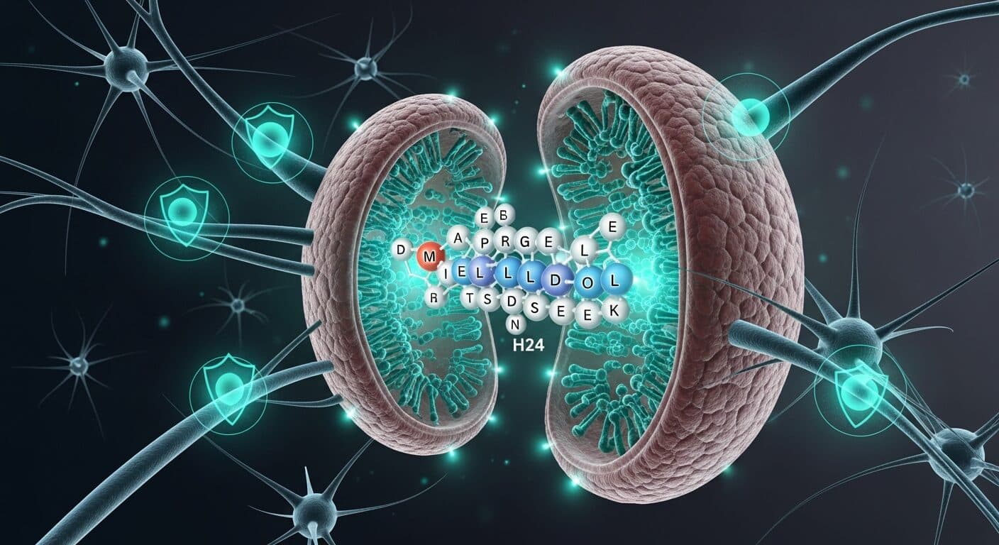
Humanin (HN) is a 24-amino-acid peptide first identified in 2001 from the surviving neurons of a brain affected by Alzheimer’s disease pathology, in a screen designed to find endogenous factors that antagonize amyloid-β-induced neuronal death [1][2]. The discovery was significant for two reasons. First, it introduced an unexpected candidate endogenous neuroprotective factor. Second, and more foundationally for molecular biology, humanin was eventually shown to be encoded by an open reading frame embedded within the mitochondrial 16S ribosomal RNA gene — making it, along with MOTS-c, a charter member of the mitochondrial-derived peptide (MDP) family [3].
For researchers working in neuroscience, mitochondrial biology, apoptosis, cardiovascular research, or aging, humanin is scientifically important because it provides a tractable experimental probe of a peptide that (a) originates in mitochondria but acts at the plasma membrane, (b) interfaces directly with the apoptotic machinery, and (c) has documented effects across multiple organ systems. This article reviews humanin’s structure, mechanisms, and the principal research areas in which it has been studied, drawing on peer-reviewed literature retrieved from PubMed.
Molecular Structure and Biochemistry
Humanin is a 24-amino-acid peptide with the sequence Met-Ala-Pro-Arg-Gly-Phe-Ser-Cys-Leu-Leu-Leu-Leu-Thr-Ser-Glu-Ile-Asp-Leu-Pro-Val-Lys-Arg-Arg-Ala [1][2]. The published sequence spans a short open reading frame within the mitochondrial 16S rRNA locus, oriented such that the peptide can be translated either from a mitochondrial or a cytoplasmic ribosome depending on which RNA population serves as template. As with MOTS-c, this dual-origin possibility has been a point of investigation for researchers studying the biogenesis of MDPs.
Key biochemical and structural features relevant to laboratory research:
- Central hydrophobic stretch (Leu9-Leu10-Leu11-Leu12) gives humanin amphipathic character and facilitates interaction with membranes and with hydrophobic binding pockets on target proteins.
- Critical residues: Structure-function studies have identified Ser14 as essential for extracellular (receptor-mediated) activity. Substitution to Gly (producing [Gly14]-humanin or HNG) markedly potentiates the neuroprotective activity of the peptide in research models [12][15]. HNG is the most widely used analog in research settings precisely because of this enhanced potency.
- Cysteine at position 8: Cys8 enables disulfide-linked dimerization, which may be relevant to some of humanin’s extracellular activities.
- Dual mechanism of action: Extracellularly, humanin engages a heterotrimeric cell-surface receptor complex composed of CNTFR/WSX-1/gp130 and can also signal through FPR2/3 and the formyl peptide receptor family. Intracellularly, humanin interacts with Bax and other Bcl-2 family proteins to inhibit mitochondrial outer membrane permeabilization [3][4].
- Evolutionary conservation: Comparative genomic analysis has shown strong synonymous codon bias across vertebrates, consistent with amino-acid-level selection pressure — robust evidence that humanin is a bona fide functional peptide rather than a translational byproduct [5].
Mechanism of Action in Research Models
Humanin operates through at least two mechanistically distinct modes, both well represented in the published research literature.
1. Extracellular, receptor-mediated neuroprotection. The original humanin research characterized it as a secreted factor that protects neurons from Alzheimer’s-disease-associated insults including amyloid-β peptides and mutant familial-AD-causing genes [1][2]. Mechanistically, extracellular humanin binds a cell-surface receptor complex (CNTFR/WSX-1/gp130) and activates downstream STAT3 signaling. Parallel studies have implicated additional receptor systems including FPR2/3. The net effect in multiple research models is suppression of caspase-dependent cell death pathways and protection of mitochondrial membrane potential under cytotoxic insults.
2. Intracellular inhibition of Bax and apoptosis. Intracellularly overexpressed or delivered humanin directly binds Bax, preventing its translocation to the mitochondrial outer membrane and the subsequent release of cytochrome c [2]. This mechanism is independent of the surface-receptor pathway and may be more relevant in research models where humanin is delivered intracellularly or expressed transgenically.
A 2018 research study in a neonatal hypoxia-ischemia rat model reported that humanin treatment attenuated the Abeta(25-35)-induced Bcl-2/Bax ratio reduction and decreased caspase-3 activity, consistent with the Bax-inhibition mechanism [15]. A 2020 peptide-chemistry study of novel humanin analogs (HUJInin, c(D-Ser14-HN)) demonstrated dose-dependent neuroprotection and myoprotection in PC12, SH-SY5Y, H9c2, and C2C12 cell lines through Erk1/2 and AKT phosphorylation and improvement of mitochondrial function [9].
Downstream signaling effects consistently reported in humanin research include:
- Caspase-3 suppression in multiple neuronal research models [6][7].
- Preservation of mitochondrial membrane potential (ΔΨm) under oxidative, ischemic, or amyloid-β insults [15].
- Reduction of cytochrome c release from mitochondria [15].
- Activation of PI3K/AKT signaling, particularly in Parkinson’s research models [13].
- Upregulation of mitochondrial biogenesis genes, suggesting an autocrine loop where humanin stimulates its own expression [13].
- Insulin sensitization at peripheral (skeletal muscle, adipose) and central (hypothalamic) levels [3][8].
Key Research Areas
1. Neuroprotection and Neurodegeneration Research
The neuroprotection research area is where humanin was first characterized and where the majority of published research still concentrates. The foundational 2004 review by Niikura and colleagues summarized the first wave of humanin research, establishing it as a potent protector against Alzheimer’s-disease-specific insults including amyloid-β toxicity in cerebrovascular smooth muscle cells and neurons [2]. A 2021 review from the same group updated the field with a comprehensive catalog of humanin’s actions in AD research contexts, including its effects on amyloid plaque accumulation and multiple intracellular anti-cell-death pathways [8].
Beyond Alzheimer’s research, humanin has been studied in Parkinson’s-disease research models. A 2023 study published in Theranostics reported that intranasal delivery of humanin rescued cell death and promoted mitochondrial function in a mouse PD model, with the peptide detected in the brain primarily via the trigeminal pathway [13]. This research established intranasal delivery as a viable administration route for research studies and demonstrated that humanin can induce its own expression via a PI3K/AKT-mediated positive feedback loop.
Other neuroprotection research areas include:
- Retinal pigment epithelium (RPE) protection against endoplasmic reticulum stress via upregulation of mitochondrial glutathione [11]. This research linked humanin to the RPE cell biology relevant to macular degeneration research.
- Neurotoxicity from silver nanoparticles in SH-SY5Y neuroblastoma cells, where humanin pretreatment protected against DNA damage, mitochondrial dysfunction, and apoptosis [14].
- PC12 cell protection against amyloid-β(25-35) toxicity using the [Gly14]-humanin (HNG) analog, with quantitative measurements of mitochondrial membrane potential, cytochrome c release, and caspase-3 activity [15].
2. Cardioprotection and Myocardial Research
A second substantial research area concerns humanin’s role in protecting cardiomyocytes from ischemia-reperfusion (I/R) injury. Multiple research groups have reported that humanin or HNG administered before, during, or at the onset of reperfusion attenuates cardiac and brain damage in preclinical I/R models.
A 2018 study in 36 male Wistar rats subjected to cardiac I/R demonstrated that HNG at 168-252 μg/kg administered during ischemia, or at 252 μg/kg at the onset of reperfusion, effectively attenuated brain mitochondrial dysfunction, tau hyperphosphorylation, amyloid-β accumulation, and apoptosis — establishing a cardiac-to-brain protective axis for humanin research [6]. A 2021 review from the Muzumdar laboratory synthesized the published evidence that humanin plays a cardioprotective role in myocardial I/R through mechanisms including autophagy modulation, ER stress reduction, cellular metabolism regulation, oxidative stress suppression, and inflammation control [10].
From a cell-biology standpoint, research on humanin analogs has shown dose-dependent myoprotection in H9c2 and C2C12 myoblast cultures exposed to doxorubicin-induced cardiotoxicity, a research-relevant model of cancer-chemotherapy-associated cardiac damage [9]. This body of work positions humanin analogs as useful experimental probes for studying the interface between mitochondrial function and cardiomyocyte survival.
3. Apoptosis and Aging Research
Humanin’s third major research domain concerns its role as a modulator of apoptosis and its decline with age in circulating levels. A 2016 study from Cobb and colleagues characterized the broader MDP family (humanin and the SHLP peptides) and reported that humanin’s circulating levels decline with age in multiple species [3]. The same study demonstrated that humanin and SHLP2 reduced apoptosis, improved mitochondrial metabolism, and enhanced adipocyte differentiation in vitro, with SHLP2 additionally acting as a central and peripheral insulin sensitizer in hyperinsulinemic-euglycemic clamp experiments [3].
For aging researchers, humanin provides a biomarker-plus-intervention framework: circulating levels can be measured as an index of mitochondrial secretory function, while exogenous administration in research models can test causal hypotheses about the age-associated decline. This dual role parallels the position of MOTS-c in the aging literature.
Stability, Storage, and Handling in the Laboratory
Humanin and its most commonly used research analog, [Gly14]-humanin (HNG), are relatively stable synthetic peptides for laboratory research use, but several handling considerations are worth noting:
- Lyophilized material should be stored at -20°C or -80°C in a desiccated environment, away from light. The peptide is hygroscopic, so vials should be equilibrated to room temperature before opening to avoid moisture condensation.
- Reconstitution solvents: Sterile water is commonly used as a primary solvent for HNG, though some research protocols use phosphate-buffered saline (PBS) or bacteriostatic water. The peptide contains one cysteine (Cys8), so reducing conditions (or inert-atmosphere storage) can help prevent unintended disulfide oxidation and dimerization for research applications where the monomeric form is desired.
- Post-reconstitution stability: Aqueous humanin solutions lose activity over time. Aliquoting into single-use volumes and freezing at -20°C to -80°C is standard research practice. Repeated freeze-thaw cycles should be avoided.
- pH: Neutral pH (7.0-7.4) is optimal for humanin stability in solution. Strongly acidic or basic conditions can accelerate degradation.
- Analog selection for research: Native humanin is functional but [Gly14]-humanin (HNG) is typically used in research applications because of its substantially greater potency — often 1,000-fold higher in neuroprotection assays [12][15]. Researchers should specify which analog they are using and report the source, purity (HPLC), and mass confirmation.
- In vivo delivery routes in published research: Intracerebroventricular, intraperitoneal, subcutaneous, intravenous, and (more recently) intranasal routes have all been used in preclinical research models [6][13]. Pharmacokinetic properties differ substantially across routes, so research protocols should be designed with attention to the intended biodistribution.
None of the above should be construed as preparation guidance for administration to humans or animals outside of an approved research protocol. These are general laboratory handling considerations.
Research Considerations and Limitations
Analog selection matters. Native humanin, HNG, and other analogs (HUJInin, c(D-Ser14-HN), colivelin) have substantially different potencies, stability profiles, and receptor selectivities [9][12]. Research results obtained with one analog should not be automatically generalized to others.
Receptor complexity. Humanin’s cell-surface signaling involves multiple receptor complexes (CNTFR/WSX-1/gp130, FPR2/3), and the relative contribution of each varies by cell type and context. Researchers should include receptor-knockdown or pharmacological antagonism controls where possible.
Intracellular vs. extracellular delivery. Because humanin has both plasma-membrane-bound (receptor-mediated) and intracellular (Bax-binding) mechanisms, experimental delivery routes can shift the dominant mechanism. Peptide uptake efficiency varies by cell type and formulation.
Mixed results across research groups. Some of humanin’s reported effects have been more reproducible than others. Researchers should cross-reference effect sizes and conditions across independent publications before designing experiments with narrow predictive windows.
Translation from preclinical to clinical research is limited. The vast majority of humanin research remains in preclinical (cell-culture and rodent) models. Human interventional data is limited, and biomarker research has been constrained by assay variability. Researchers should exercise appropriate caution in interpreting translational claims.
Frequently Asked Research Questions
Q1: What is the difference between humanin and [Gly14]-humanin (HNG)?
HNG is a single-residue analog in which the serine at position 14 is replaced with glycine. This substitution dramatically increases the neuroprotective potency of humanin in research models, by as much as three orders of magnitude in some assays [12]. HNG is the most widely used analog in neuroprotection research, while native humanin is more commonly used in studies focused on biomarker measurement or mechanistic dissection of receptor binding [15].
Q2: How does humanin differ mechanistically from MOTS-c?
Both are mitochondrial-derived peptides encoded within mitochondrial rRNA regions (16S for humanin, 12S for MOTS-c). Humanin acts predominantly through cell-surface receptors (CNTFR/WSX-1/gp130, FPR2/3) and intracellular Bax inhibition, with a primary biological activity as an anti-apoptotic and neuroprotective factor [1][2][3]. MOTS-c acts predominantly through intracellular AMPK activation and nuclear translocation, with its primary biological activities in metabolic and exercise-related research [3]. Their receptor biology and signaling pathways are largely non-overlapping, even though they share an anatomical origin in the mitochondrial ribosome.
Q3: What are the best-characterized positive and negative controls for humanin experiments?
In neuroprotection research models, positive controls typically include well-established neuroprotective agents such as N-acetylcysteine or specific pathway inhibitors (e.g., caspase inhibitors for Bax-pathway experiments). Negative controls include scrambled humanin sequences or mutant variants in which critical residues (Ser14, Cys8) are substituted to abolish activity [2][12]. Vehicle-only treatment provides the baseline for comparison.
Q4: Can humanin cross the blood-brain barrier in research models?
Published research suggests that intravenously or peripherally administered humanin has limited BBB penetration, which is why intracerebroventricular and intranasal delivery routes have been favored for CNS-focused research [13]. The 2023 Theranostics study demonstrated that intranasal humanin distributes primarily via the trigeminal pathway, providing an alternative to invasive CNS delivery for research purposes [13].
Q5: How is circulating humanin typically measured in plasma samples?
Published humanin biomarker research has relied on ELISA-based immunoassays developed against the full-length peptide or specific epitopes. Reported plasma concentrations vary substantially across studies due to assay differences, matrix effects, and sample-handling variability [3][8]. Researchers planning biomarker work should validate their assay against synthetic humanin standards and report recovery and inter-assay variability.
References
According to PubMed, the following peer-reviewed articles were used as primary sources for this research overview. DOI links are provided where available.
- Niikura T. Humanin and Alzheimer’s disease: The beginning of a new field. Biochim Biophys Acta Gen Subj. 2022;1866(1):130024. PMID: 34626746. DOI
- Niikura T, Chiba T, Aiso S, Matsuoka M, Nishimoto I. Humanin: after the discovery. Mol Neurobiol. 2004;30(3):327-40. PMID: 15655255. DOI
- Cobb LJ, Lee C, Xiao J, et al. Naturally occurring mitochondrial-derived peptides are age-dependent regulators of apoptosis, insulin sensitivity, and inflammatory markers. Aging (Albany NY). 2016;8(4):796-809. PMID: 27070352. DOI
- Gong Z, Goetzman E, Muzumdar RH. Cardio-protective role of Humanin in myocardial ischemia-reperfusion. Biochim Biophys Acta Gen Subj. 2022;1866(2):130066. PMID: 34896254. DOI
- Gruschus JM, Morris DL, Tjandra N. Evidence of natural selection in the mitochondrial-derived peptides humanin and SHLP6. Sci Rep. 2023;13(1):14110. PMID: 37644144. DOI
- Kumfu S, Charununtakorn ST, Jaiwongkam T, Chattipakorn N, Chattipakorn SC. Humanin Exerts Neuroprotection During Cardiac Ischemia-Reperfusion Injury. J Alzheimers Dis. 2018;61(4):1343-1353. PMID: 29376862. DOI
- Matsunaga D, Sreekumar PG, Ishikawa K, et al. Humanin Protects RPE Cells from Endoplasmic Reticulum Stress-Induced Apoptosis by Upregulation of Mitochondrial Glutathione. PLoS One. 2016;11(10):e0165150. PMID: 27783653. DOI
- Niikura T. Humanin and Alzheimer’s disease: The beginning of a new field. Biochim Biophys Acta Gen Subj. 2022;1866(1):130024. PMID: 34626746. DOI
- Gilon C, Gitlin-Domagalska A, Lahiani A, et al. Novel humanin analogs confer neuroprotection and myoprotection to neuronal and myoblast cell cultures exposed to ischemia-like and doxorubicin-induced cell death insults. Peptides. 2020;134:170399. PMID: 32889021. DOI
- Gong Z, Goetzman E, Muzumdar RH. Cardio-protective role of Humanin in myocardial ischemia-reperfusion. Biochim Biophys Acta Gen Subj. 2022;1866(2):130066. PMID: 34896254. DOI
- Matsunaga D, Sreekumar PG, Ishikawa K, et al. Humanin Protects RPE Cells from Endoplasmic Reticulum Stress-Induced Apoptosis by Upregulation of Mitochondrial Glutathione. PLoS One. 2016;11(10):e0165150. PMID: 27783653. DOI
- Jin H, Liu T, Wang WX, et al. Protective effects of [Gly14]-Humanin on beta-amyloid-induced PC12 cell death by preventing mitochondrial dysfunction. Neurochem Int. 2010;56(3):417-23. PMID: 19941922. DOI
- Kim KH. Intranasal delivery of mitochondrial protein humanin rescues cell death and promotes mitochondrial function in Parkinson’s disease. Theranostics. 2023;13(10):3330-3345. PMID: 37351170. DOI
- Gurunathan S, Jeyaraj M, Kang MH, Kim JH. Mitochondrial Peptide Humanin Protects Silver Nanoparticles-Induced Neurotoxicity in Human Neuroblastoma Cancer Cells (SH-SY5Y). Int J Mol Sci. 2019;20(18):4439. PMID: 31505887. DOI
- Jin H, Liu T, Wang WX, et al. Protective effects of [Gly14]-Humanin on beta-amyloid-induced PC12 cell death by preventing mitochondrial dysfunction. Neurochem Int. 2010;56(3):417-23. PMID: 19941922. DOI
Disclaimer: All products sold by CertaPeptides are intended for laboratory research use only. Not for human or veterinary use. Not for consumption. This article is provided for scientific and educational purposes and does not constitute medical advice, dosing guidance, or an endorsement of any particular use. Researchers are responsible for complying with all applicable institutional, national, and international regulations governing the acquisition, handling, and study of peptide research compounds.