⚠️ For Research Purposes Only — This article discusses follistatin and the follistatin-344 isoform as laboratory research compounds. The content is intended for scientific and educational review only. The peptide is not for human consumption and not intended to diagnose, treat, cure, or prevent any disease. All studies referenced describe preclinical investigations in cell culture or animal models.
Introduction
Follistatin is a secreted, cysteine-rich glycoprotein that functions as a high-affinity binding protein and antagonist for several members of the transforming growth factor-beta (TGF-β) superfamily, most notably activins and myostatin. In vertebrates, follistatin is expressed as multiple isoforms generated by alternative splicing of the single FST gene. The two principal isoforms in human biology are follistatin-288 (FS-288) and follistatin-315 (FS-315); a third major isoform, follistatin-344 (FS-344), represents the primary mRNA transcript that encodes the full-length protein prior to proteolytic processing and is widely referenced in preclinical gene delivery research and muscle physiology studies.
In preclinical skeletal muscle research, follistatin and follistatin-derived constructs have been investigated as tools to probe myostatin signaling, activin receptor pathway biology, and the feasibility of local versus systemic modulation of muscle mass in rodent models. According to PubMed, transgenic mice expressing follistatin in skeletal muscle exhibit dramatic increases in muscle mass comparable to those observed in myostatin-null animals (Lee and McPherron 2001, DOI).
This article reviews the biochemistry of follistatin, its molecular interactions with myostatin and activin, the major research paradigms in which follistatin-based tools have been used, and the practical considerations for handling and interpreting follistatin research.
Molecular Structure and Biochemistry
Follistatin is encoded by the FST gene, which produces multiple transcripts through alternative splicing. The main isoforms are:
- Follistatin-288 (FS-288): A shorter isoform that retains strong affinity for cell-surface heparan sulfate proteoglycans via its heparin-binding domain, yielding largely tissue-localized activity. FS-288 and engineered FS-288-Fc constructs have been used extensively to probe localized, rather than systemic, modulation of muscle mass in rodents (Castonguay et al. 2018, DOI).
- Follistatin-315 (FS-315): A longer isoform with an acidic C-terminal extension that reduces heparin binding and supports systemic circulation.
- Follistatin-344 (FS-344): Represents the full-length precursor transcript (344 residues), and is the form commonly referenced in adeno-associated virus (AAV) gene delivery research where the unprocessed polypeptide is introduced to muscle for sustained local expression of functional follistatin.
Structurally, follistatin contains an N-terminal domain (ND) followed by three characteristic cysteine-rich follistatin domains (FSD1, FSD2, FSD3). Together these domains form the ligand-binding surfaces that wrap around myostatin or activin dimers and block interactions with type I and type II receptors (Saitoh et al. 2020, DOI).
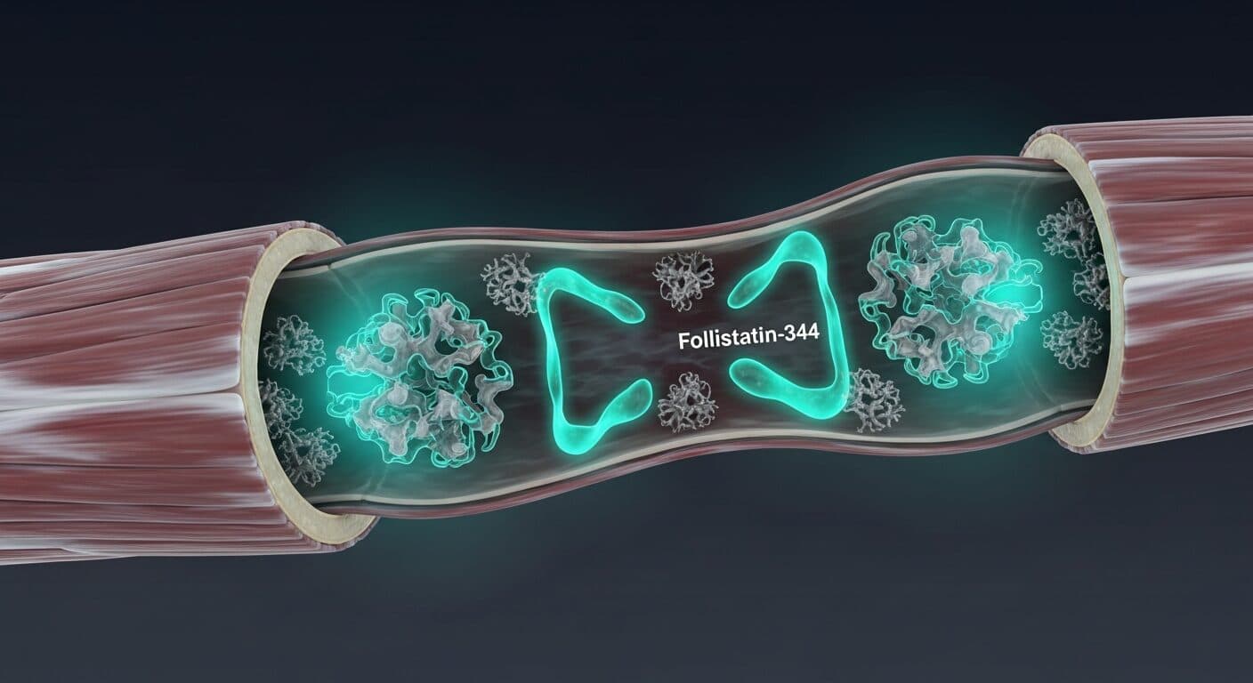
Follistatin binds myostatin and activins with nanomolar affinity. A central biochemical feature is that two follistatin molecules encircle a single TGF-β ligand dimer, sterically preventing engagement of ActRIIB/ActRIIA and downstream SMAD2/3 signaling. This molecular mechanism explains the potent negative effect of follistatin on myostatin activity in cell-based assays and in vivo rodent work (Lee and McPherron 2001, DOI).
Mechanism of Action in Research Models
Myostatin Pathway Inhibition
Myostatin is a TGF-β family ligand produced primarily by skeletal muscle that acts as a negative regulator of muscle mass. It binds ActRIIB (and to a lesser extent ActRIIA), recruits ALK4/ALK5, and triggers SMAD2/3 phosphorylation, which suppresses myogenic differentiation and protein synthesis and promotes protein breakdown. A foundational biochemical study demonstrated that purified myostatin C-terminal dimer binds ActRIIB and ActRIIA, and that follistatin can inhibit this binding in vitro; transgenic mice overexpressing follistatin, the myostatin propeptide, or a dominant-negative ActRIIB in skeletal muscle all showed dramatic muscle mass increases comparable to those observed in myostatin knockout animals (Lee and McPherron 2001, DOI). This parallel phenotype across distinct blockade strategies established follistatin as a robust molecular tool for interrogating myostatin biology.
Comprehensive reviews summarize the biology of myostatin and its regulators, including follistatin, in skeletal muscle growth and wasting contexts (Esposito et al. 2021, DOI; Wagner 2005, DOI; Chen and Lee 2016, DOI).
Activin and Broader TGF-β Ligand Binding
Follistatin also binds activin A and activin B and, depending on the isoform and construct, can modulate GDF11 and other TGF-β family ligands. This multi-ligand capture is central to follistatin’s biology because activins and myostatin share downstream receptors. Research using an engineered follistatin-288-Fc fusion protein demonstrated high-affinity binding to activin A, activin B, myostatin (GDF8), and GDF11 with functional neutralization in cell-based reporter assays (Castonguay et al. 2018, DOI). The same study showed that intramuscular administration of FST288-Fc in mice induced dose-dependent local muscle growth without affecting surrounding or contralateral muscles, contrasting with systemically active ActRIIB-Fc, which produced generalized effects.
Heparin Binding and Tissue Localization
A key biochemical feature of the FST-288 isoform is its heparin-binding property, which restricts its diffusion and localizes activity to tissues expressing heparan sulfate proteoglycans. This makes FST288-based constructs particularly useful research tools for probing the consequences of localized TGF-β ligand blockade, as opposed to systemic inhibition (Castonguay et al. 2018, DOI).
Follistatin-Derived Peptides
More recent medicinal chemistry work has sought to identify minimal follistatin-derived peptides that retain myostatin-selective inhibitory activity. One group identified a 14-mer peptide (DF-3) from the N-terminal domain of follistatin that inhibited myostatin signaling in a luciferase reporter assay without affecting activin A or TGF-β1, and that increased skeletal muscle mass in mice when delivered intramuscularly (Saitoh et al. 2020, DOI). These data reinforce the biological relevance of the follistatin ND in myostatin binding and offer a template for studying isolated myostatin pathway inhibition in rodents.
Key Research Areas
1. Skeletal Muscle Physiology and Growth
The headline research finding for follistatin in muscle biology is its ability to expand muscle mass when expressed or administered in rodent models, paralleling the phenotype of myostatin loss. This has made follistatin a cornerstone investigative tool for dissecting the molecular architecture of muscle protein turnover, myofiber hypertrophy, and the interplay between satellite cell activity and differentiated myofiber signaling (Lee and McPherron 2001, DOI; Chen and Lee 2016, DOI; Wagner 2005, DOI).
2. Preclinical Models of Muscle Wasting and Dystrophinopathies
Because myostatin inhibition is a rational strategy for counteracting muscle atrophy, follistatin and related tools have been studied in preclinical models of sarcopenia, muscular dystrophy, and cachexia. In dysferlinopathy patient cohorts, serum myostatin and follistatin have been evaluated as candidate biomarkers for disease monitoring; baseline myostatin correlated with function and MRI measures, though changes over time did not track with individual patient trajectories, suggesting myostatin’s role as a disease monitoring biomarker is limited while its biology as a drug target remains of interest (Moore et al. 2023, DOI). In community-dwelling older women and men, plasma follistatin, myostatin, and GDF11 have been correlated with muscle function measures, highlighting the relevance of the myostatin/follistatin axis to aging muscle research (Fife et al. 2018, DOI).
3. Body Composition, Metabolism, and Exerkine Research
Beyond raw hypertrophy, follistatin sits within the broader “exerkine” framework, where exercise-responsive circulating factors influence muscle, adipose, and metabolic tissues. Reviews of exercise-responsive cytokines note follistatin-family members among the exerkine axis relevant to osteoarthritis and metabolic research (Jia et al. 2023, DOI). A recent review on weight loss-associated lean mass preservation emphasizes the myostatin/activin/follistatin/inhibin system as a central node for understanding lean mass dynamics, with pharmacological strategies that target activins or myostatin being actively explored in preclinical and clinical research (Stefanakis et al. 2024, DOI).
4. AAV Gene Delivery and Follistatin-344 Constructs
Follistatin-344 is the mRNA isoform most commonly referenced in adeno-associated virus (AAV) gene therapy research, where the unprocessed 344-residue polypeptide is expressed locally in muscle to drive sustained follistatin output. Preclinical studies have demonstrated that intramuscular injection of AAV-FS344 vectors can produce dose-dependent muscle hypertrophy in rodents over weeks to months. The rationale for using the full-length precursor is that endogenous proteolytic processing generates both FS-288 and FS-315 variants within the target tissue, maximizing the functional versatility of the expressed protein. Researchers working with gene-delivered follistatin systems need to carefully validate vector titer, transgene expression kinetics, and downstream muscle biomarker responses (e.g., myofiber cross-sectional area, p-SMAD2/3 levels, and GDF8/activin A neutralization in muscle lysates).
5. Comparative Pharmacology: Follistatin, ActRIIB-Fc, and Myostatin Antibodies
The preclinical muscle hypertrophy literature includes several distinct classes of myostatin pathway modulators: follistatin and follistatin fusion proteins, decoy ActRIIB-Fc receptors, myostatin-selective antibodies (e.g., trevogrumab, domagrozumab in pharmaceutical nomenclature), activin-selective antibodies (e.g., garetosmab), bimagrumab-class ActRII blockers, and small-molecule myostatin inhibitors. Each class has a different ligand selectivity profile and different tissue-distribution characteristics, which shape both efficacy and off-target biology in rodent models (Stefanakis et al. 2024, DOI; Chen and Lee 2016, DOI). Follistatin’s broad ligand capture (myostatin, activin A, activin B, GDF11) makes it a useful tool for interrogating the combined contribution of these ligands to muscle phenotype, whereas myostatin-selective reagents are better suited for narrow mechanism studies.
Exerkine and Systemic Signaling Roles
Follistatin is emerging as an important “exerkine” — a circulating factor released during exercise that mediates cross-tissue signaling between muscle and distant organs. Reviews of exerkines in osteoarthritis and metabolic health highlight follistatin family proteins alongside myokines like irisin, IL-6, and metrnl as mediators of exercise-induced adaptation, with both protective and maladaptive effects depending on context (Jia et al. 2023, DOI). In aging research, plasma follistatin, myostatin, and GDF11 levels correlate with muscle function measures in older adults, suggesting that the myostatin/follistatin axis is responsive to age-related changes in body composition and physical activity (Fife et al. 2018, DOI). These data position follistatin as a biomarker of interest in addition to its role as a mechanistic tool.
Stability, Storage, and Handling in the Laboratory
Follistatin and follistatin-based constructs are glycoproteins and peptides with specific handling needs:
- Lyophilized protein storage: Typically stored at −20 °C or −80 °C in sealed, desiccated containers. Once reconstituted, short-term storage at 2–8 °C for 24–72 hours is common.
- Reconstitution: Sterile water or PBS is standard. Low-binding polypropylene tubes help limit surface adsorption losses.
- Carrier proteins: 0.1% BSA in working dilutions is commonly used to reduce adsorptive losses and preserve activity during long incubations.
- Freeze-thaw cycles: Follistatin constructs should be aliquoted to minimize repeated freeze-thaw cycles, which can cause aggregation and loss of ligand-binding activity.
- Heparin sensitivity: FST-288 isoforms bind heparin and heparan sulfate; tissue-level distribution in vivo is correspondingly restricted relative to FST-315 or Fc-fusion variants lacking heparin affinity (Castonguay et al. 2018, DOI).
- Identity and activity QA: Routine quality control includes SDS-PAGE, mass spectrometry identification, and bioactivity assays such as myostatin-responsive luciferase reporters or ActRIIB binding.
- Endotoxin testing: Recombinant follistatin prepared in bacterial systems must be tested for endotoxin, as LPS contamination can confound downstream cytokine, NF-κB, and cell viability readouts. Lysate assays (LAL) or recombinant factor C assays are the standard approach.
- Sterility and pyrogenicity: For in vivo rodent studies, sterile-filtered preparations are standard. Working dilutions should be prepared immediately before injection to minimize adsorptive losses and microbial contamination.
Analytical Methods for Follistatin Research
Researchers studying follistatin in preclinical models rely on a layered set of analytical tools. SDS-PAGE and Western blot distinguish the main follistatin isoforms by size and can be combined with anti-FST antibodies for detection in tissue lysates. ELISA platforms measure total circulating follistatin in rodent plasma, though most commercial kits do not distinguish between FS-288 and FS-315. Functional assays include myostatin-responsive SMAD-luciferase reporter cell lines (e.g., HEK293 transfected with CAGA-luc), ActRIIB binding assays by SPR or ELISA, and direct measurement of p-SMAD2/3 in muscle tissue by Western blot. MRI-based muscle volume and fat fraction measurements have been used to track follistatin-related interventions in dystrophy models (Moore et al. 2023, DOI). Combining biochemical readouts (p-SMAD, transcriptomics) with morphological readouts (fiber cross-sectional area, MRI) and functional readouts (grip strength, rotarod) gives the strongest mechanistic conclusions.
Research Considerations and Limitations
Researchers designing follistatin-centered studies should carefully consider:
- Ligand promiscuity. Native follistatin binds multiple TGF-β ligands (myostatin, activin A, activin B, GDF11, and others), so experimental effects cannot always be cleanly attributed to myostatin inhibition. Reporter assays with defined ligands and follistatin-derived peptides with sharper selectivity can help dissect these contributions (Saitoh et al. 2020, DOI; Castonguay et al. 2018, DOI).
- Systemic vs. local pharmacology. FST-288, FST-315, FST-344, and engineered Fc fusions have different tissue distribution profiles because of heparin binding and proteolytic processing. Results from one construct do not automatically transfer to another.
- Proteolytic stability in circulation. Some follistatin constructs undergo rapid proteolysis in circulation, which can restrict activity to intramuscular injection sites (Castonguay et al. 2018, DOI).
- Compensatory biology. Chronic myostatin/activin pathway suppression triggers compensatory responses in other TGF-β family ligands and receptors, and long-term phenotypes may differ from acute ones (Esposito et al. 2021, DOI).
- Species differences. Rodent muscle biology does not always translate directly to larger animal models; comparative work in livestock species highlights useful nuances for interpretation (Chen and Lee 2016, DOI).
Historical Context and Discovery
Follistatin was originally isolated in the 1980s from ovarian follicular fluid as a factor that suppresses follicle-stimulating hormone (FSH) secretion, and its name reflects this first characterized function. Subsequent work in the 1990s revealed that follistatin binds and neutralizes activin, which explained its effect on FSH regulation through the activin-driven FSH signaling axis. The connection to muscle biology came later, after the discovery of myostatin (GDF8) as a negative regulator of muscle mass in a landmark 1997 study by Se-Jin Lee and Alexandra McPherron. When the same group demonstrated that follistatin binds and inhibits myostatin, and that transgenic overexpression of follistatin in skeletal muscle recapitulates the hypertrophic phenotype of myostatin-null animals, the follistatin/myostatin/muscle axis became one of the most productive areas of translational skeletal muscle research (Lee and McPherron 2001, DOI).
Since then, follistatin has attracted attention as a research tool for studying muscle biology, a biomarker for muscle-wasting conditions, and a template for therapeutic strategies aimed at preserving lean mass in catabolic states. The development of engineered follistatin fusion proteins, gene therapy vectors expressing FS-344, and follistatin-derived peptides illustrates the creative ways researchers have exploited the core follistatin architecture for specific experimental applications.
Frequently Asked Research Questions
Q1: What distinguishes follistatin-344 from follistatin-288 and follistatin-315?
FST-344 refers to the full-length precursor polypeptide (344 residues), while FST-288 and FST-315 arise from alternative splicing and proteolytic processing. FST-288 has strong heparin-binding activity and is tissue-localized; FST-315 circulates more widely due to an acidic C-terminal extension that reduces heparin affinity.
Q2: Does follistatin only inhibit myostatin?
No. Follistatin binds multiple TGF-β superfamily ligands including activin A, activin B, myostatin (GDF8), and GDF11 with high affinity, as demonstrated in biochemical and cell-based assays (Castonguay et al. 2018, DOI).
Q3: What receptor does myostatin signal through?
Myostatin signals primarily through activin receptor type IIB (ActRIIB) and, to a lesser extent, ActRIIA, recruiting ALK4/ALK5 and activating SMAD2/3 (Lee and McPherron 2001, DOI).
Q4: Are there follistatin-derived peptides that are selective for myostatin?
Yes. A 14-mer peptide named DF-3, derived from the N-terminal domain of follistatin, selectively inhibited myostatin without affecting activin A or TGF-β1 in reporter assays and increased muscle mass after intramuscular injection in mice (Saitoh et al. 2020, DOI).
Q5: What readouts are used in preclinical follistatin studies?
Common readouts include muscle wet weight, fiber cross-sectional area, grip strength, SMAD2/3 phosphorylation in muscle lysates, myostatin-responsive luciferase reporter activity in cell lines, and MRI-derived muscle volume and fat fraction measurements (Moore et al. 2023, DOI).
References
- Lee SJ, McPherron AC. Regulation of myostatin activity and muscle growth. Proceedings of the National Academy of Sciences USA. 2001;98(16):9306-9311. DOI (PMID: 11459935).
- Castonguay R, Lachey J, Wallner S, et al. Follistatin-288-Fc Fusion Protein Promotes Localized Growth of Skeletal Muscle. Journal of Pharmacology and Experimental Therapeutics. 2019;368(3):435-445. DOI (PMID: 30563942).
- Saitoh M, Takayama K, Hitachi K, et al. Discovery of a follistatin-derived myostatin inhibitory peptide. Bioorganic & Medicinal Chemistry Letters. 2020;30(3):126892. DOI (PMID: 31874826).
- Esposito P, Picciotto D, Battaglia Y, Costigliolo F, Viazzi F, Verzola D. Myostatin: Basic biology to clinical application. Advances in Clinical Chemistry. 2022;106:181-234. DOI (PMID: 35152972).
- Wagner KR. Muscle regeneration through myostatin inhibition. Current Opinion in Rheumatology. 2005;17(6):720-724. DOI (PMID: 16224249).
- Chen PR, Lee K. INVITED REVIEW: Inhibitors of myostatin as methods of enhancing muscle growth and development. Journal of Animal Science. 2016;94(8):3125-3134. DOI (PMID: 27695802).
- Moore U, Fernández-Simón E, Schiava M, et al. Myostatin and follistatin as monitoring and prognostic biomarkers in dysferlinopathy. Neuromuscular Disorders. 2023;33(2):199-207. DOI (PMID: 36689846).
- Fife E, Kostka J, Kroc Ł, et al. Relationship of muscle function to circulating myostatin, follistatin and GDF11 in older women and men. BMC Geriatrics. 2018;18(1):200. DOI (PMID: 30165829).
- Stefanakis K, Kokkorakis M, Mantzoros CS. The impact of weight loss on fat-free mass, muscle, bone and hematopoiesis health: Implications for emerging pharmacotherapies aiming at fat reduction and lean mass preservation. Metabolism. 2024;161:156057. DOI (PMID: 39481534).
- Jia S, Yu Z, Bai L. Exerkines and osteoarthritis. Frontiers in Physiology. 2023;14:1302769. DOI (PMID: 38107476).
Closing Remarks for Researchers
Follistatin and its isoforms, including the full-length follistatin-344 precursor form, sit at the center of one of the most productive areas of contemporary muscle biology research. The peptide’s ability to sequester multiple TGF-β family ligands with high affinity, combined with the availability of engineered fusion proteins, AAV-delivered constructs, and minimal follistatin-derived peptides, gives investigators a rich palette of tools for interrogating myostatin and activin signaling in rodent and cell-based models. The field’s ongoing translation of follistatin biology into candidate pharmacological interventions for muscle wasting, sarcopenia, and cachexia underscores both the scientific importance and the therapeutic promise of this research axis. For laboratory investigators, follistatin-based reagents provide a reliable way to suppress myostatin pathway signaling and generate a clear muscle hypertrophy phenotype, enabling mechanistic dissection of downstream pathways, transcriptional responses, and cellular adaptations. As always, all follistatin-related research materials discussed in this article are intended solely for laboratory use in controlled preclinical settings.
References retrieved from PubMed. All DOI links point to primary sources.
Disclaimer: All products sold by CertaPeptides are intended for laboratory research use only. Not for human or veterinary use. Not for consumption. Not for human consumption. For laboratory research only.